
Head CT scan
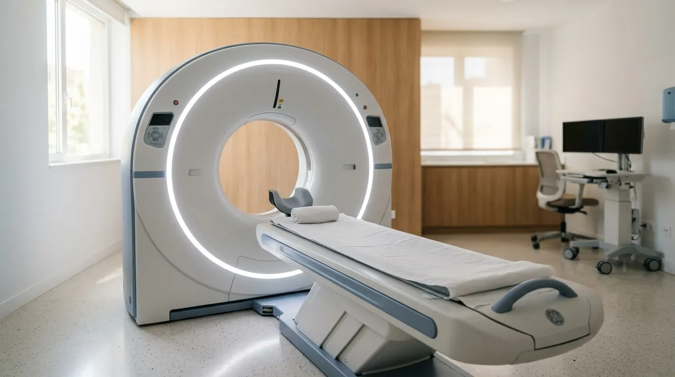
Head CT scan: computed tomography of the head for head injuries, headaches with warning signs, sinuses and intracranial conditions.
What a head CT scan is
Computed tomography takes many X-ray images from different angles and combines them into slices that show the inside of the head in great detail. It's used to assess the skull bone, brain tissue, the ventricles, the sinuses and the temporal bone.
It's a medical test, different from the so-called «dental CT» — cone beam tomography used to plan implants —, which is optimised for bone and teeth and isn't suitable for examining the brain.
First things first
There are situations where you don't book an appointment for a CT scan: go to A&E instead. A sudden, extremely intense headache that peaks within seconds; a head injury with loss of consciousness or repeated vomiting; sudden weakness, slurred speech or vision loss; or a seizure need immediate care, and that's where it's decided what test is needed and when. This page is for information and doesn't replace that assessment.
When it's indicated
- Head injury, especially with loss of consciousness, vomiting, older age or blood-thinning medication. Not every knock to the head needs a CT scan: there are clinical criteria to decide.
- Headache with warning signs: sudden onset, fever with a stiff neck, neurological deficit, a change in pattern in someone who already gets headaches, or a first episode from age 50 onwards.
- Suspected intracranial condition: bleeding, a mass, or hydrocephalus.
- Examining the sinuses in recurring sinusitis or before nasal surgery.
- Bone assessment of the ear and temporal bone in selected cases of hearing loss or vertigo.
When it isn't needed
Most headaches don't need an imaging test. If the neurological examination is normal and there are no warning signs, a CT scan adds no information and does add radiation; what helps then is a good medical history.
And a CT scan isn't always the best test for the head: for brain tissue, the back of the brain, the auditory nerve or a stroke in its first hours, MRI sees much more. CT wins on speed and availability — that's why it's the test used in emergencies — and for examining bone.
What the test involves
You lie on your back on a table that moves inside an open ring: it isn't a long tunnel, so claustrophobia is far less of an issue than with MRI. It takes a few minutes and you only need to stay still. It doesn't hurt and, if no contrast is used, needs no preparation or fasting.
If contrast is used
For some indications, iodine-based contrast is given through a vein. In that case you'll be asked about previous reactions to contrast, your kidney function and the medication you take — some diabetes medications need a specific schedule around it — and you'll be asked to fast for a few hours. During the injection it's normal to feel warmth and a metallic taste for a few seconds.
Radiation, pregnancy and children
The dose from a head CT scan is much higher than an X-ray: roughly what's received from natural background radiation over several months. That isn't a reason to refuse the test when it's indicated — the benefit of a correct diagnosis is far greater —, but it is why it isn't done routinely or «just for peace of mind».
Always mention if you are or might be pregnant. In children the criteria are stricter, the technique is adjusted to their size and, wherever possible, a test without radiation is preferred.
The result
The scan is interpreted and reported by a radiologist, and the images are provided digitally so you can take them to your specialist. It's worth being clear that the report isn't the diagnosis: it describes what's seen, and it's your doctor who puts it alongside your symptoms and examination to decide what it means.
Frequently asked questions
Is a CT scan the same as an MRI?
No. The CT scan uses X-rays and takes slices within a few minutes inside an open ring, so claustrophobia is far less of a problem. MRI uses magnetic fields, takes longer and is a tunnel. Each one answers different questions.
Will I be given contrast?
Only for certain indications. When iodinated contrast is given intravenously, you are asked beforehand about allergies, kidney function and medication, and fasting for a few hours is required. If no contrast is used, no preparation is needed.
When is a head CT scan indicated?
After a head injury, especially with loss of consciousness, vomiting or if you take anticoagulants; with a headache showing warning signs; to study the paranasal sinuses in recurrent sinusitis; and to assess the ear in selected cases.
When should you have a CT scan and when an MRI?
The CT scan is fast and better for bone, acute bleeding and trauma: it is the emergency test. The MRI shows soft tissue in far more detail —brain, spinal cord, tendons— and uses no radiation, but it takes longer and is not always available. What you are looking for determines which one is indicated.
When is a brain CT scan necessary?
After a head injury with loss of consciousness, vomiting or while on anticoagulants; with a sudden, extremely intense headache or one with neurological signs; with suspected stroke; and to study the paranasal sinuses or the ear in selected cases. It is not indicated for an ordinary headache without warning signs.