
3D dental scan (DentaScan / CBCT)
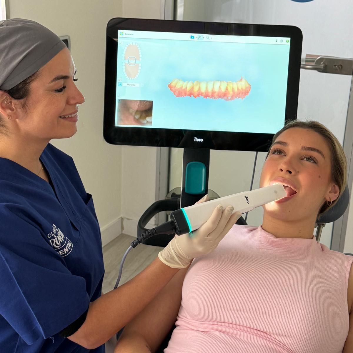
A three-dimensional radiographic study of the upper and lower jaw: it measures the height, width and density of the bone before a dental implant is placed.
What it is and what it adds
A dental X-ray is flat: it shows height and width, but not depth. Two crossing roots appear on top of one another and there is no way of telling which one is in front. The dental scan reconstructs the area in three dimensions and then allows it to be sliced in any plane.
That changes the surgeon's work: they can measure the available bone in real millimetres and see exactly where the structures that must be respected are —the inferior alveolar nerve, the maxillary sinus, the neighbouring roots— before operating, rather than during.
You will see this test under several names: dental scan, DentaScan, dental CT or CBCT. Today it is carried out with cone beam computed tomography (CBCT), a machine specific to the maxillofacial area that makes a single turn around the head in a matter of seconds.
When it is needed
- Planning for dental implants: how many, what size, in which position and at what angle, and whether a bone graft is required.
- Extraction of wisdom teeth when the panoramic X-ray suggests that the roots are close to the nerve.
- Root canal treatment in complicated cases: canals that cannot be located, unusual anatomy, retreatments and suspected root fracture.
- Impacted teeth, cysts and other lesions of the jaws.
- Assessment of the jaw joint and surgical planning.
When it is not needed
The 3D scan is not the first test and it does not replace the panoramic X-ray. International guidelines say so plainly: it is not used routinely and it is not requested if a lower-dose test is enough to make the decision. For most check-ups, straightforward orthodontic cases and fillings, it adds nothing that cannot already be seen.
It is justified when there is a specific question that a flat image cannot answer; almost always, how much bone there is and how far away the nerve lies.
What the test is like
Standing or sitting, with your head steadied and biting on a small support. It takes a few seconds. You have to remove all metal —earrings, piercings, hairpins, removable dentures— and stay still: movement spoils the reconstruction here more than it does on a standard X-ray.
It does not hurt, no contrast is injected, there is no need to fast and you do not go into any tunnel: the machine is open and you are standing up, so claustrophobia is not usually a problem.
How much radiation it involves
This is the point that tends to be explained worst. The dose of a dental scan is higher than that of a panoramic X-ray —considerably higher, and the larger the area explored the higher it is— and clearly lower than that of a medical CT scan of the same region.
In other words: it is a low-dose test compared with a hospital CT scan, not compared with a dental X-ray. That is why the field is adjusted to the smallest area needed, and why it is not requested «while we're at it». If you are pregnant or think you may be, please tell us beforehand.
In children and adolescents
The same criterion, applied more strictly: growing tissue is more sensitive to radiation, so in under-18s the 3D scan is reserved for situations where it really will change the treatment —an impacted canine, a lesion, a developmental anomaly— and always with the smallest possible field.
What you receive
As well as the images, the study is supplied in digital format so the case can be planned with the dentist's or surgeon's software, including guided surgery. It is usually requested together with the panoramic X-ray, which remains the overall view.
If your dentist has recommended a dental scan, or you have been referred by another professional with the test already requested, book an appointment and tell us what you have been asked for.
Frequently asked questions
When is a 3D dental scan essential?
Before placing implants, to measure the bone actually available; with wisdom teeth whose roots are close to the nerve; in complex root canals or retreatments; and to study impacted teeth, cysts or the jaw joint.
Is it the same as a hospital CT scan?
It is the same idea —three-dimensional imaging— but with a dental cone beam unit, focused on the jaws and with a considerably lower radiation dose than a conventional medical CT scan. The test takes seconds and is done sitting or standing.
Can I take the file to another dentist?
The study is provided in digital format, which is what allows the case to be planned with the dentist's or surgeon's software, including guided surgery. Ask for it at the clinic and it will be given to you together with the report.