
Dental radiology
Panoramic X-ray, intraoral X-ray, 3D scanner and lateral cephalogram, at the clinic itself, in the same visit.
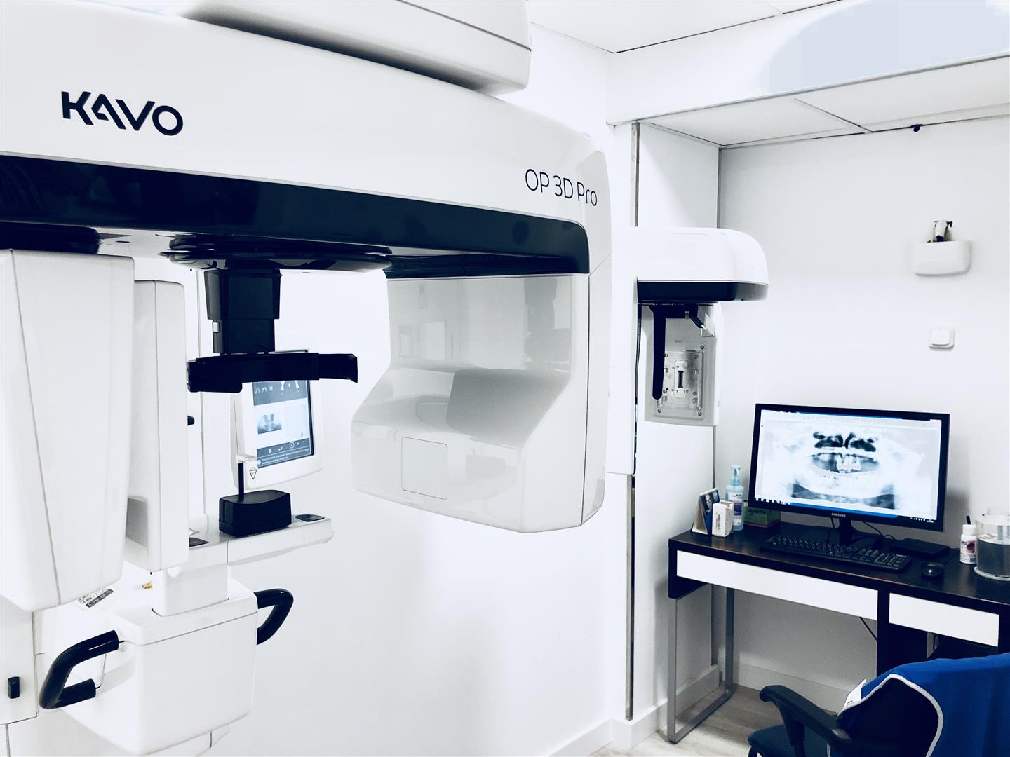
Dental diagnosis starts with the image
Almost no treatment is decided by looking inside the mouth alone. To know the state of the bone, where the nerve runs, whether a root is infected or how far a tooth has shifted, you need to see what can't be seen. That's what dental radiology is for.
Which tests we do
- Orthopantomogram — the panoramic X-ray: the whole mouth in one image, teeth, upper and lower jaw. It's the general view almost every study starts with.
- Intraoral X-ray — the close-up detail of one or two teeth, with the sensor inside the mouth. It's the one that finds decay hidden between teeth and root problems.
- 3D dental scan (DentaScan / CBCT) — the three-dimensional study of the upper and lower jaw: height, width and bone density, millimetre by millimetre. Also the jaw joint, the sinuses and the airway, for what a panoramic X-ray can't resolve.
- Lateral cephalogram — the profile X-ray used to measure facial proportions and angles.
- Intraoral scanner — the digital 3D copy of your mouth. It's here because it's an imaging diagnostic tool, but it's worth knowing that it isn't an X-ray and it emits no radiation: it's an optical camera that replaces impression putty.
When you book, we confirm which clinic your test will be done at.
When each one is needed
The test isn't chosen at random: each treatment needs to see something different.
- First visit and check-ups. Orthopantomogram, plus an intraoral X-ray when a specific tooth needs a closer look.
- Orthodontic assessment. Panoramic plus lateral cephalogram: the first shows which teeth there are and their condition, the second how the jaws fit together.
- Root canal treatment and pain with no clear cause. Intraoral X-ray, and a 3D study when the canal anatomy is complex.
- Wisdom teeth and oral surgery. Panoramic to see the position, and a scan when the tooth is close to the nerve.
What 3D dental radiology adds
A standard X-ray flattens something that has volume into a single plane: two roots that cross come out overlapping, with no way to tell which is in front. The cone beam CT (CBCT) rotates around the head and reconstructs the area as a volume, which can then be sliced along any plane.
That allows the bone where an implant will go to be measured in real millimetres, the dental nerve to be located, a maxillary sinus assessed or an oddly shaped root understood before it's touched. In implantology and surgery, it's the difference between planning and improvising.
How much radiation you receive
Dental tests are among the lowest-dose in the whole of medical radiology, and our equipment is digital: the sensor needs considerably less exposure than old-style film. Even so, every test is requested because it will change a clinical decision, not as routine.
During the test you wear protection and staff stay with you: it takes seconds, standing or sitting, with no discomfort and no need to prepare beforehand.
🔴 If you are pregnant or think you might be, say so before the test. You don't need to wait for us to ask.
How to book a test
Usually your dentist will indicate it during your consultation, and it's done there and then or you're booked in for it. If you've been referred by another professional with a specific test requested, we can also do it for you: book an appointment and tell us what's been requested.
Frequently asked questions
What X-rays are taken at a dental clinic?
Intraoral X-rays, taken with a sensor inside the mouth and giving maximum detail of a single tooth; the panoramic, which shows both complete arches; the lateral cephalogram for orthodontics; and the 3D scan, which provides volume and real measurements for surgery and implants.
How much radiation do I get at the dentist?
Very little. With today's digital equipment, dental doses are among the lowest in all of medical radiology and protective measures are used. Even so, they are only requested when they add information for the diagnosis or the treatment.
I am pregnant, can I have a dental X-ray?
Always say so before the test. Many dental X-rays can be postponed without any risk to the treatment; when they are essential —an acute infection, for example— they are taken with protection and limiting the examination to what is necessary.
How is a dental X-ray taken?
It depends on the type: for an intraoral X-ray a small sensor is placed inside the mouth next to the tooth and you are asked to bite and stay still for a second; for a panoramic you bite on a positioner and the equipment rotates around your head for a few seconds; for a 3D scan the rotation is similar but it captures volume. In all of them you have to remove metal and keep still.
What can't a pregnant woman have done at the dentist?
You can and should go to the dentist during pregnancy: pregnancy gingivitis is very common and a cleaning is indicated. What is postponed, unless urgent, are X-rays and scheduled treatments that can wait, and certain drugs are avoided. Always tell us that you are pregnant and how many weeks along you are.
Areas within this specialty