
Intraoral scanner
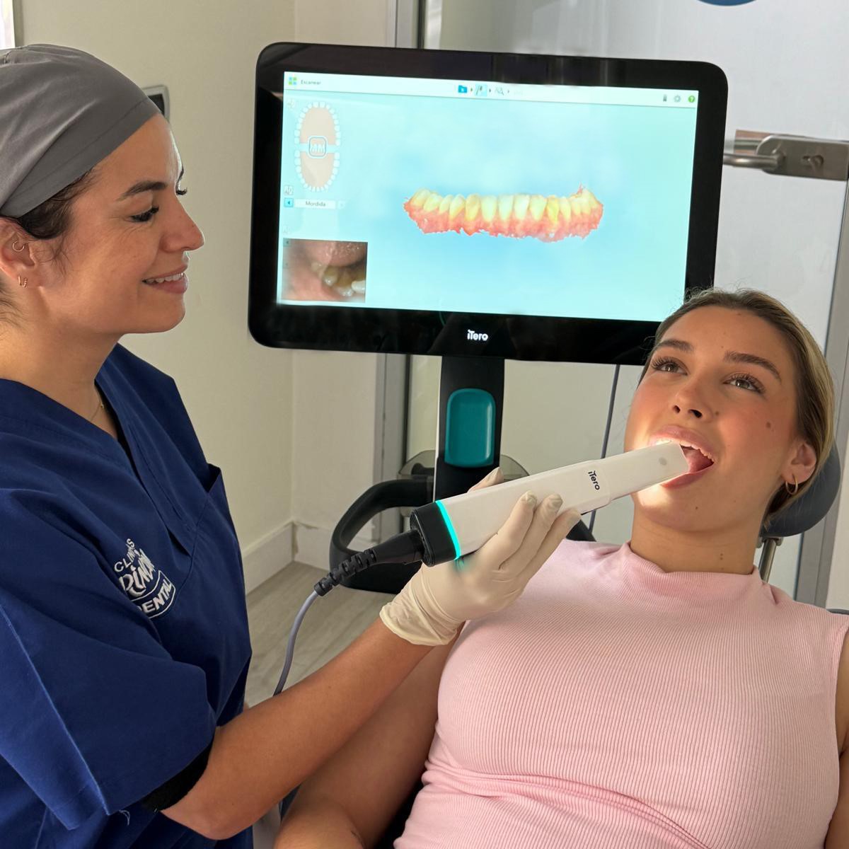
The iTero scanner reproduces the mouth in 3D in just a few minutes: it replaces paste impressions and lets you see the treatment on screen during the same visit.
What an intraoral scanner is
It is a camera about the size of a thick pen that travels over the teeth and gums and gradually builds on the screen a three-dimensional model of your mouth. It replaces the impressions of old: the trays full of paste that you had to put up with for several minutes and that made many people gag.
It is worth saying clearly, because it causes confusion: it is not an X-ray and it emits no radiation. It is light, not X-rays. It appears in this section because it is diagnostic imaging, but it has nothing in common with the other radiology tests.
What it is used for
- Design and manufacture of crowns, bridges and prostheses.
- Night guards for bruxism.
- Planning of implants: the surface model is combined with the 3D dental scan, which supplies the bone, in order to design guided surgery.
- Monitoring of tooth wear or gum recession, comparing scans taken on different dates.
What it cannot see
The intraoral scanner copies surfaces: what can be seen inside the mouth. It does not pass through anything, so it does not show the inside of the tooth, or the roots, or the bone, or decay hidden between two teeth. X-rays are still needed for that.
It is a test that complements radiology, not an alternative to it: if anyone offers you a scan as a substitute for an X-ray, it is not one.
What it is like for the patient
You open your mouth and the camera travels over both arches and your bite. It takes a few minutes, it does not hurt, there are no needles or pastes, and it can be paused and resumed whenever you want: if you need to swallow, take a break or have a gag reflex, it stops and then continues. That is why it is usually tolerated far better than a traditional impression.
It requires no preparation beforehand and no fasting.
What the treatment gains
Fewer repeats —if one area does not come out well, only that part is scanned again, not the whole mouth—, a file that reaches the laboratory immediately and usually shortens the timescales, and a model that is kept on file: if something has to be redone later on, there is no need to take another impression.
And an advantage that is not technical but does count: the model is shown enlarged on the screen, so you can look at your own mouth while it is explained to you what is going on and what is being proposed. Decisions are easier to understand with the image in front of you.
Would you like to know whether your case can be planned this way? Book an appointment and we will assess it.
Frequently asked questions
Does the intraoral scanner emit radiation?
No. It is an optical camera: it passes over the teeth and builds a three-dimensional model from images, without any kind of radiation. It can be repeated as often as needed, even to compare how the mouth changes over time.
Does it replace impressions taken with putty?
In most cases yes, and it is one of the best pieces of news for anyone with a gag reflex. It can also be paused and resumed, and if one area comes out poorly only that part is rescanned instead of repeating the whole impression.