
Lateral cephalometric X-ray
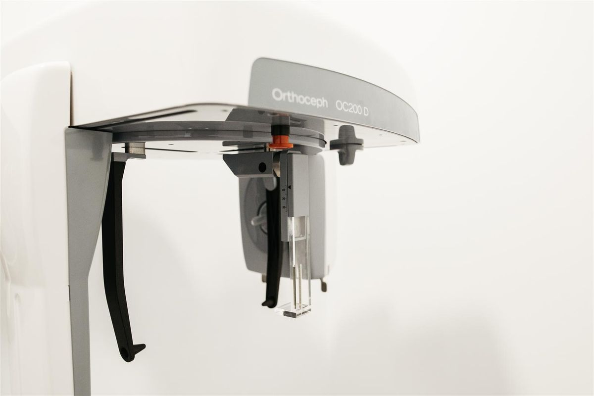
A profile X-ray taken from two metres away to avoid distortion: it is the basis of the orthodontic study because it measures the bones and proportions of the face.
What it is and why it has that name
It is an X-ray of the head taken in profile, with the patient always held in the same position and with the X-ray tube well away —a metre and a half or more— from the head. That distance is the reason for the name («tele-») and it is not a whim: the further away the source is, the more parallel the rays arrive and the less the image is distorted.
Thanks to that, what is measured on the image matches the real measurement, and it can be compared with that of another patient or with that of the same patient a year later. Without that standardisation, the X-ray would be useful for looking at, but not for measuring.
What it is for: cephalometry
The cephalometric studyis carried out on this image: specific anatomical points are marked and angles and distances are calculated that describe how the maxilla, the mandible, the teeth and the profile of the face relate to one another. It is what makes it possible to answer a question that cannot be settled by eye: whether the teeth are badly positioned on well-placed bones, or whether the mismatch is in the bones themselves.
- Initial orthodontic study when the relationship between the jaws has to be assessed.
- Monitoring of progress during treatment and at the end of it.
- Planning together with the surgeon when the correction is not only dental but also skeletal.
- Follow-up of facial growth in children and adolescents under treatment.
- Assessment of the upper airway space and of adenoid size in selected cases.
When it is not needed
Not every orthodontic treatment needs a cephalometric X-ray. When the case is straightforward and the problem is only the position of the teeth, the panoramic X-ray and the scan of the mouth are enough. Requesting it routinely for everyone starting orthodontics is not justified: it is done when the answer is going to change the treatment plan.
How it is done
Standing, with the head held by two supports placed in the ears —so the position is always identical—, looking straight ahead, with the teeth in contact and the lips relaxed. Earrings, glasses, hairpins and any metal must be removed.
It takes a few seconds, it does not hurt, nothing is placed in the mouth and it requires no preparation or fasting.
Radiation, pregnancy and children
It is one of the lowest-dose X-rays there is: below what you receive from natural background radiation in a single day. Most of the people who have it are children and adolescents, and precisely for that reason the criterion is applied more strictly: only if it is going to change a decision, and with the field adjusted to the bare minimum.
If you are pregnant or think you may be, tell us before the test.
What you receive
The image is supplied in digital format so the orthodontist can trace the cephalometric analysis on it. It is usually requested together with the panoramic X-ray: the panoramic shows which teeth are there and how they are; the cephalometric shows how the bones supporting them fit together. Together they make up the basic radiological study for most orthodontic treatments.
If you would like to start with an assessment, book an appointment: we will tell you which tests your case needs, without taking any for granted.
Frequently asked questions
Why does the orthodontist ask for a lateral X-ray?
Because the cephalometric analysis is done on it: anatomical points are marked and angles and distances are measured that show how the upper jaw, the lower jaw, the teeth and the profile relate to one another. Without that analysis an orthodontic treatment cannot be properly planned.
Is it repeated when treatment finishes?
It is usually repeated to compare the final situation with the initial one and to check that the planned movements have been achieved. In growing patients it may also be repeated during treatment to assess how the jaw is developing.
How do you position yourself for this X-ray?
Standing up, with the head held by two supports at the ears that always ensure the same position, looking straight ahead, with the teeth touching and the lips relaxed. That repeatability is what makes it possible to compare X-rays taken on different dates.