
Consejos
Published on
MRI results with findings: are they the cause of my pain?
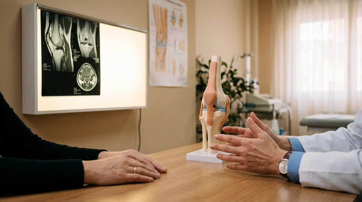
MRI results often show herniated discs, protrusions or meniscal wear in people with no pain. How your report is matched with your physical examination.
Not necessarily. MRI scans very often find disc protrusions, meniscal wear or tendinosis in people who have no pain at all, and these are more common the older you are. A finding appearing in your MRI results doesn't mean it is the source of what you're feeling: that is decided by relating the image to your history and to the physical examination at your appointment.
Why MRI results with findings don't always explain your pain
Magnetic resonance imaging is a very sensitive test: it shows discs, menisci, tendons and cartilage in detail. That same sensitivity means it picks up changes from the passing of the years and from use that don't have to hurt, just as grey hair doesn't hurt.
Studies in people with no symptoms show this clearly:
- Lower back. Among people with no back pain, disc wear is already common at age 20 and almost universal at 80. Protrusions also appear at all ages and become more common over the years.
- Knee. In middle-aged and older people, a meniscal tear is a common finding, and more than half of those who have one had noticed no pain, aching or stiffness in the previous month.
- Shoulder. Changes in the rotator cuff tendons, those surrounding the joint, also become more common with age, whether or not the people who have them have symptoms.
None of this means your MRI doesn't matter. It means that, on its own, it isn't enough to know what is causing your pain.
Common words in an MRI report
The radiologist describes everything they see, whether or not it's relevant to your case, and often without knowing your symptoms in detail. These are some common expressions and what they usually describe:
- Disc dehydration or degeneration: the disc has lost water and height over the years. This is one of the most common findings from middle age onwards.
- Disc bulge or protrusion: the disc sticks out slightly beyond its edge. It may not touch any nerve.
- Disc herniation or extrusion: part of the disc comes out further. It becomes important when what you feel fits with the nerve that could be affected.
- Degenerative meniscal tear or meniscopathy: wear of the meniscus, often along with changes in the cartilage.
- Tendinosis or tendinopathy: changes in the structure of the tendon, common in the shoulder or the Achilles tendon.
- Degenerative or arthritic changes: wear of the joint, to a varying degree.
- Joint effusion or bone oedema: fluid inside the joint or in the bone. Depending on the context, these can be mild or point to an injury that should be looked into.
These are descriptions, not diagnoses. The diagnosis is made by whoever puts the image together with what is happening to you.
What decides whether a finding is causing your pain: the examination
In a trauma and orthopaedics appointment, the question isn't what shows up on the MRI, but which part of what shows up explains what's happening to you. Three things count in answering it:
- Your history: how the pain started, whether there was a knock or a twist, which movements make it worse, whether it wakes you at night and whether it spreads to another area, and where to.
- The examination: movement, strength, stability, manoeuvres that load a particular tendon or meniscus and, if the problem is your back, the sensation and reflexes in your legs.
- The match: whether the image and the examination point to the same place. A herniated disc on the left side is a poor explanation for pain running down the right leg; meniscal wear doesn't carry the same weight with a truly locked knee as with years of vague pain.
That's why, with degenerative meniscal tears, surgery isn't usually the first step, as explained in the article on knee arthroscopy and meniscal injuries.
When a finding does change the treatment
The image carries more weight when it fits with the examination and when its result can change the decision. For example:
- An injury after a clear trauma, such as a torn ligament or a torn meniscus that locks the knee.
- Compression of a nerve that matches pain running down the arm or leg and the loss of strength or sensation found on examination.
- Pain that doesn't improve despite proper treatment for a reasonable time, or when surgery is being considered.
In all other cases, the usual approach is to treat what the examination finds: exercise and physiotherapy, adjusting the load, pain control and, in some cases, injections. The pages on shoulder pain and sports injuries explain how these problems are investigated and when an MRI is reserved for them.
How to bring your MRI to your appointment
- The report and the images, on a disc or with the download link if you were given one. The images show what the report summarises.
- Previous MRI scans or X-rays of the same area, even from another centre: comparing them shows whether anything has changed.
- A brief note of when the pain started, what makes it worse and what you've tried: medication, rest, physiotherapy or previous injections.
- Your questions in writing. Two useful ones: ‘Does this finding fit with what's happening to me?’ and ‘Would another test change what we're going to do?’
For lower back pain, routine imaging tests aren't recommended: they are reserved for when the result can change the treatment. Repeating an MRI out of worry tends to add findings, not answers.
Signs not to wait
Whatever your report says, go to A&E or call 112 if you develop:
- Increasing loss of strength in one leg, or sudden difficulty walking.
- Loss of control of your urine or bowels, or numbness in the genital area or between your buttocks.
- Back pain with a fever, or after a fall or a heavy blow.
- Inability to put weight on or move a joint after an injury, or a visible deformity.
Also see a doctor without delay if the pain is new, severe and doesn't ease with rest, or if you have a history of cancer.
Interpreting your MRI with the specialist
MRI results with findings are valuable information when someone puts them in context. If you have a report that worries you and pain that won't go away, the trauma and orthopaedics clinic can help you put the report alongside the examination and decide which treatment makes sense in your case.
Frequently asked questions
Shouldn't the radiologist say whether the finding is important?
The radiologist describes in detail everything they see in the image, and often doesn't know about your examination. Deciding which finding explains the pain is up to the doctor who examines you and knows your history.
Do I need to repeat the MRI if I'm still in pain?
Not routinely. It's considered if your symptoms change, a new problem appears on examination or surgery is being considered and the image could change the decision. The specialist decides after examining you.
With a disc protrusion or herniated disc on my MRI, can I exercise?
For back pain, staying active and exercising are part of the recommended treatment. Which exercise and how much load are adjusted after the examination, and you should stop and see a doctor if you develop increasing loss of strength or tingling.
Is an MRI done at another centre any use?
Generally, yes. Bring the report and, if you can, the images, along with previous tests of the same area. Comparing MRI scans from different dates helps to show whether anything has changed.
Based on reliable sources
Prepared by our team from the references listed below. This is general information and does not replace a consultation.
Article pending review by one of our doctors.
References (4)
- Brinjikji W et al. Systematic literature review of imaging features of spinal degeneration in asymptomatic populations. AJNR Am J Neuroradiol. 2015;36(4):811-6 Open
- Englund M et al. Incidental meniscal findings on knee MRI in middle-aged and elderly persons. N Engl J Med. 2008;359(11):1108-15 Open
- Teunis T et al. A systematic review and pooled analysis of the prevalence of rotator cuff disease with increasing age. J Shoulder Elbow Surg. 2014;23(12):1913-21 Open
- NICE NG59. Low back pain and sciatica in over 16s: assessment and management (recomendaciones 1.1.4-1.1.6 sobre imagen) Open