
Echocardiogram
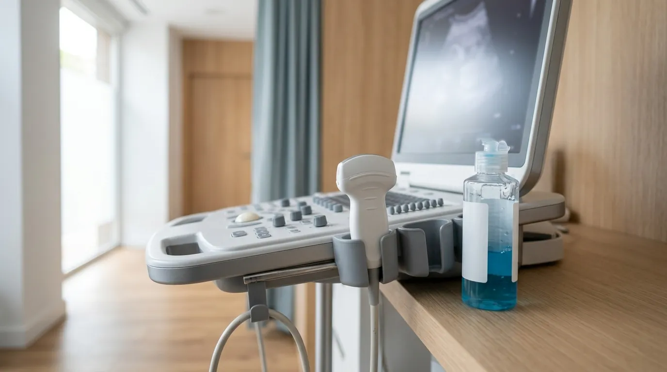
Transthoracic Doppler echocardiogram: an ultrasound of the heart that assesses the valves, walls and pumping function. No radiation, no preparation.
What an echocardiogram shows
An echocardiogram uses ultrasound — the same as any ultrasound scan — to see the heart beating in real time. Doppler is also used to study how blood flows inside it. A single test assesses:
- The pumping strength of the left ventricle.
- The state of the valves: whether they close properly or are narrowed.
- The thickness of the walls and the size of the chambers.
- The pericardium, the membrane surrounding the heart.
When it's requested
- A murmur picked up during an examination.
- Breathlessness, unusual tiredness or swollen legs.
- Years of high blood pressure, to see how it's affected the heart.
- Follow-up of a known valve condition or heart failure.
- Investigating palpitations or chest pain, alongside an electrocardiogram.
What the test involves
You lie on your left side, with your chest uncovered. A gel is applied and the probe is placed at different points. It takes between fifteen and thirty minutes, doesn't hurt, uses no radiation and needs no preparation: no fasting, no stopping medication. You can carry on as normal afterwards.
Afterwards
The cardiologist explains what they've seen and links it to your symptoms. From there comes the plan: discharge, regular monitoring, adjusting treatment or, if needed, referral for a more complex test.
Frequently asked questions
Does an echocardiogram involve radiation?
No. It uses ultrasound, the same as any other scan, so it can be repeated as often as needed and is also performed during pregnancy. Nor does it need contrast in most cases or any prior preparation: no fasting and no stopping medication.
What exactly can be seen?
The heart beating in real time: how strongly the left ventricle pumps, the state of the valves —whether they close properly or are narrowed—, the thickness of the walls, the size of the chambers and the pericardium. With Doppler, how the blood flows inside is studied as well.
How long does it take and can I carry on as normal afterwards?
Between fifteen and thirty minutes. You lie on your left side with your chest uncovered, gel is applied and the probe is placed at different points. It does not hurt. When it is over you can leave and carry on as normal; the cardiologist explains what they have seen and what the plan is.
Which conditions are detected on an echocardiogram?
Valve disease —stenosis and regurgitation—, heart failure due to poor pumping, cardiomyopathies, hypertrophy caused by high blood pressure, pericardial effusion, congenital heart disease and the consequences of a previous heart attack. It is the reference test for assessing the structure and function of the heart.
What should I do before an echocardiogram?
Nothing special: it requires no fasting and no stopping your treatment. Come in clothing that is easy to remove from the waist up, because the chest has to be uncovered, and bring previous reports and the list of your medication. The test lasts between fifteen and thirty minutes and afterwards you can carry on as normal.
Questions and comments
Ask the specialist who reviews this page or leave us a comment. Everything is reviewed before it is published.
To write, first confirm your email: we will send you a code.
Content reviewed by Dr David Grande Prada